



HEPASKIN – From Skin Tumour Screening to Oncological Treatment and Surgical Reconstruction
FotoFinder technology, medical caravans, sentinel lymph node biopsy and reconstructive surgery – components of an integrated approach to patients with skin tumours in the Romania–Serbia cross-border region.
Skin cancer is a major public health concern, and early detection followed by appropriate treatment can decisively influence patient prognosis. Malignant melanoma, one of the most aggressive forms of skin cancer, as well as basal cell and squamous cell carcinomas, require a complex medical approach that integrates prevention, diagnosis, staging, surgical treatment and long-term follow-up.
In this context, the HEPASKIN – RORS00307 project, implemented under the Interreg IPA Romania–Serbia Programme, promotes the development of an integrated care model for patients with skin tumours through the use of modern diagnostic technologies, the organisation of medical caravans and collaboration between specialists from different medical fields.
FotoFinder – modern technology for the early detection of skin tumours
One of the key investments made within the HEPASKIN project is the FotoFinder system, a high-performance digital videodermoscopy device designed for the assessment and monitoring of skin lesions.
This technology allows detailed examination of pigmented and non-pigmented lesions, identification of suspicious dermoscopic features and digital documentation of skin changes.
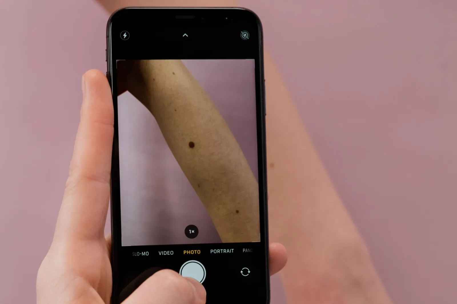
A major advantage is the ability to monitor how lesions evolve over time by comparing images obtained at successive examinations. This is particularly important for patients with numerous pigmented naevi, a personal or family history of melanoma, or other risk factors.
Computer-aided analysis tools can support the dermatologist in assessing suspicious lesions, without replacing clinical examination and the specialist’s interpretation.
FotoFinder thus contributes to the early identification of suspicious lesions, to the selection of patients who require further investigation and to reducing the risk of delayed diagnosis.
Important: the definitive diagnosis of a skin tumour is established by histopathological examination, when indicated.
HEPASKIN caravans – access to screening for communities in the cross-border region
An essential component of the project is the organisation of medical screening caravans in Romania and Serbia.
Through these activities, dermatological assessment is brought closer to the population, especially in communities where access to specialised medical services is limited.
In Romania, project activities have included localities such as Șag and Variaș in Timiș County, as well as Drobeta-Turnu Severin in Mehedinți County.
The medical caravans help identify people with suspicious skin lesions, provide medical recommendations and refer patients for further investigation and treatment.
At the same time, these activities contribute to public health education by promoting sun protection, skin self-examination and early medical consultation when suspicious changes appear.
Melanoma diagnosis and the importance of sentinel lymph node biopsy
Malignant melanoma is a form of skin cancer with significant metastatic potential, for which early diagnosis and accurate staging are essential in establishing the treatment strategy.
How the diagnosis is established
When a lesion is suspected of being melanoma, the diagnosis is usually established by excisional biopsy with histopathological examination. The histopathology report provides essential information, including Breslow thickness, the presence or absence of ulceration and other prognostically relevant features.
What is the sentinel lymph node?
Sentinel lymph node biopsy plays an important role in the staging of certain patients with melanoma.
The sentinel lymph node is the first lymph node, or first group of lymph nodes, to receive lymphatic drainage from the area of the primary tumour. Identifying and examining it makes it possible to detect possible nodal micrometastases that are not revealed by routine clinical or imaging examination.
Sentinel lymph node biopsy is particularly valuable for regional staging and for assessing melanoma prognosis, helping to determine subsequent treatment and follow-up.
When is the procedure indicated?
The indication for this procedure is established individually, based on tumour thickness, ulceration, clinical stage and oncology guideline recommendations. The procedure is considered mainly for melanomas with a Breslow thickness greater than 0.8 mm or for certain thinner, ulcerated melanomas, and is not required in every case.
How the sentinel lymph node is identified
The sentinel lymph node can be identified using lymphatic mapping techniques with radioactive tracers and/or dyes, including indocyanine green fluorescence in experienced centres with appropriate protocols.
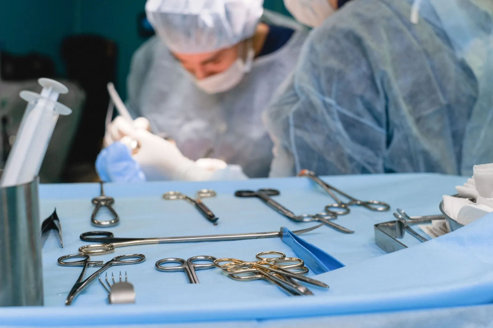
Key point: sentinel lymph node biopsy is not a method for diagnosing the primary skin tumour, but an important step in melanoma staging in selected patients.
The multidisciplinary oncology board – treatment decisions tailored to each patient
Modern treatment of melanoma and complex skin tumours requires collaboration between several medical specialties.
To this end, the multidisciplinary oncology board, also known as the Tumour Board, plays a central role in assessing cases and establishing the treatment plan.
Depending on the particularities of each case, the board may include:
- dermatologists;
- general surgeons;
- plastic surgeons;
- medical oncologists;
- pathologists;
- radiologists;
- radiation oncologists.
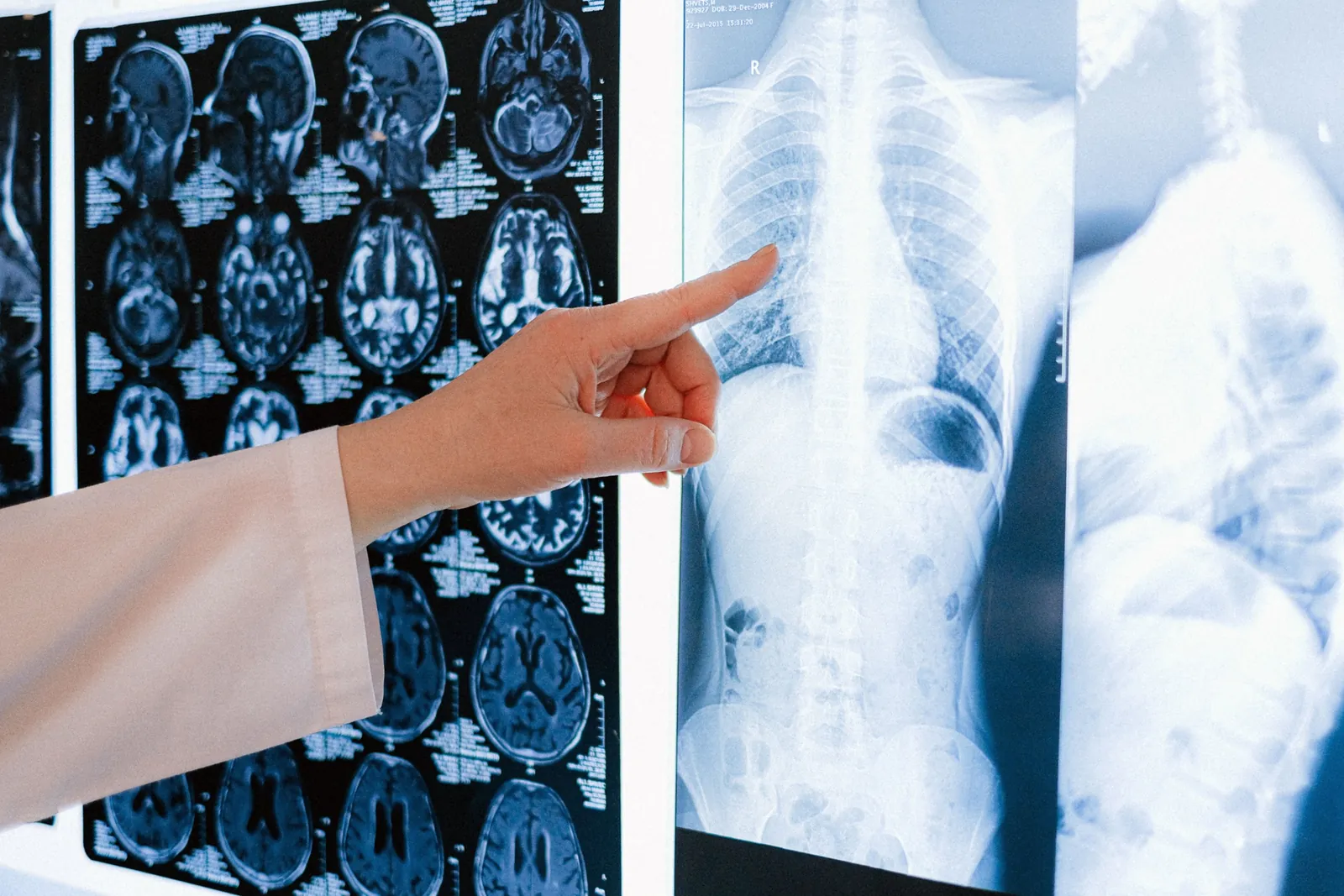
Multidisciplinary analysis makes it possible to correlate clinical, dermoscopic, histopathological and imaging information in order to establish an individualised treatment plan.
In the case of melanoma, the board may review:
- the need for sentinel lymph node biopsy;
- the indication for wide local excision;
- the appropriateness of adjuvant or neoadjuvant systemic therapies;
- the oncological follow-up strategy.
For advanced skin tumours or those located in difficult anatomical areas, multidisciplinary assessment allows surgery and reconstruction to be planned in line with oncological principles while preserving function and appearance.
The main advantage of the multidisciplinary oncology board is a coherent treatment approach, grounded in the characteristics of the disease and the particularities of the patient.
Plastic and reconstructive surgery – repairing defects after skin tumour excision
Surgical treatment is a fundamental component in the management of most localised skin tumours.
Oncological excision must aim for complete removal of the tumour, respecting the safety margins recommended for the histological type and stage of the disease.
In certain situations, particularly with large tumours or those located on the face, scalp, neck or extremities, excision may leave significant tissue defects that cannot be closed directly without tension or without affecting neighbouring anatomical structures.
In these cases, plastic and reconstructive surgery plays an essential role in restoring skin continuity, preserving function and achieving an appropriate aesthetic result.
Reconstruction techniques
Post-excision defects can be reconstructed using different techniques, adapted to the size, depth and location of the defect:
- Direct closure, when the characteristics of the defect allow it.
- Local flaps – advancement, rotation or transposition – which use tissue adjacent to the defect.
- Free skin grafts, split-thickness or full-thickness, to cover selected defects.
- Regional or pedicled flaps, for complex defects.
- Free microsurgical flaps, when extensive reconstruction with transfer of vascularised tissue is required.
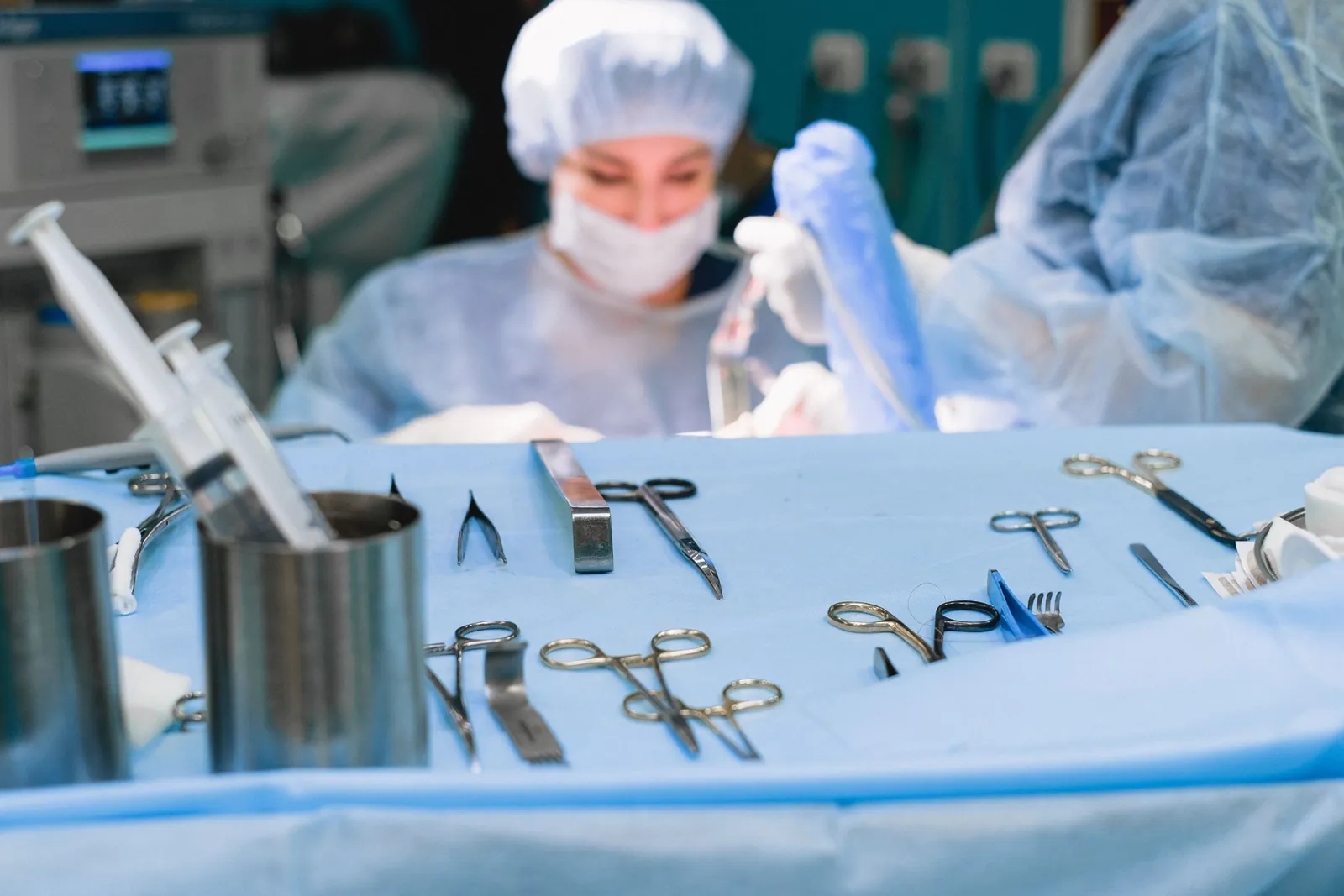
The choice of reconstructive technique aims to achieve stable coverage, protect deep structures, reduce complications and preserve the function of the affected anatomical region.
Planning the reconstruction
For tumours located on the face, reconstruction is of additional importance, as it must take into account the function of the eyelids, nose, lips and other anatomical structures, as well as the aesthetic and psychological impact on the patient.
Reconstruction planning must be correlated with oncological radicality, certainty of the resection margins and any complementary oncological treatments. In certain situations, definitive reconstruction may be deferred until the margin status has been clarified.
By integrating plastic and reconstructive surgery into the treatment of skin tumours, the therapeutic approach goes beyond simply removing the tumour, also aiming at functional recovery and the patient’s return to everyday life.
A complete care pathway – from screening to follow-up
One of the key principles promoted by the HEPASKIN project is continuity of patient care.
The complementary stages in the management of skin tumours are:
- dermatological assessment;
- digital documentation with FotoFinder;
- histopathological diagnosis;
- oncological staging;
- discussion of complex cases by the multidisciplinary board;
- surgical treatment, including reconstruction when needed.
Not every patient needs all of these stages. The care pathway is adapted to the diagnosis, the stage of the disease and the clinical indications.
A cross-border skin tumour registry
Developing a cross-border registry dedicated to skin tumours can help document cases, monitor patients and evaluate the outcomes of screening and treatment.
Through the organised collection of medical information, in compliance with personal data protection rules, the registry can become a valuable tool for research, quality-of-care assessment and the development of future prevention programmes.
HEPASKIN – an integrated approach to skin cancer
The HEPASKIN project highlights the importance of combining prevention, modern technology, medical expertise and cross-border collaboration.
The medical caravans increase access to screening, the FotoFinder system supports the identification and monitoring of suspicious lesions, sentinel lymph node biopsy enables regional staging of melanoma in selected patients, and the multidisciplinary oncology board facilitates an individualised treatment strategy.
Complementing these stages, plastic and reconstructive surgery offers solutions for covering defects resulting from tumour excision, contributing to patients’ functional and aesthetic recovery.
The central message of the HEPASKIN project is that the modern management of skin tumours is not limited to identifying a suspicious lesion, but involves an integrated care pathway – from screening and early diagnosis to staging, oncological treatment, reconstruction and long-term follow-up.
By developing medical infrastructure and strengthening collaboration between specialists in Romania and Serbia, the project helps lay the groundwork for medical services that are more accessible, better coordinated and focused on patients’ needs.
Project: HEPASKIN – RORS00307
Programme: Interreg IPA Romania–Serbia
Institution: “Victor Babeș” University of Medicine and Pharmacy, Timișoara
Project manager: Prof. Univ. Dr. Blidișel Iulian Alexandru Ciprian
Information and promotional material on the activities of the HEPASKIN project.
